
From Theory to Practice:
How ITMO Scientists Created a Wireless Compact Device for Breast Screening
Breast cancer is one of the most widespread oncological diseases and one of the most dangerous for women overall: 685,000 died of it last year alone. Doctors admit that there are no efficient ways to treat breast cancer at terminal stages. That’s why it’s important to detect the tumor and treat it as early as possible. In this case, there is a good chance of complete recovery.
Magnetic resonance imaging (MRI) is an efficient non-invasive method for early diagnostics. However, this procedure is quite expensive and requires special equipment, which makes it hardly accessible to the public. Scientists from all over the world are working on increasing the efficiency of MRI. Specialists from ITMO’s School of Physics and Engineering have been engaged in this task for eight years. Fundamental research started in 2013 and since then, the scientists have made a series of discoveries and experiments. As a result, they came up with MetaCoil – a wireless, ergonomic, compact (and aesthetic!) device for breast screening. Here’s how the project developed from a theoretical concept to a ready-to-use device.
How MRI works

MRI makes it possible to detect tumors and metastases at early stages. During the procedure, the patient is lying inside a big transmitting coil (a coil is a specialized antenna for the excitation and reception of a magnetic resonance signal). It forms a powerful radio-frequency field and the magnets located around the coil create a strong and constant magnetic field (in medical scanners it’s around 1-3 Tesla). The intersection of these fields causes a magnetic resonance in protons inside the human body. The protons get excited and begin to release energy as radio waves, and that is what the coil detects. Different organs and tissues have different proton density, so the signal varies – visually, this can be seen as contrasting fragments on resulting images.

However, the protons’ response is so weak that it’s hard to distinguish it among the noise. That’s when receiver coils developed specifically for scanning various body parts come in handy. For example, there are receiver coils for the head, body, back, legs, arms, and so on. These coils collect the magnetic resonance signal and provide images with a better quality and brightness.
MRI is the most efficient tool for diagnostics but it isn’t perfect. The resulting images often have defects, blind zones, and unclear fragments. That’s why clinicians aren’t always able to detect a disease at its early stages.
MRI is the most efficient tool for diagnostics but it isn’t perfect. The resulting images often have defects, blind zones, and unclear fragments. That’s why clinicians aren’t always able to detect a disease at its early stages.

Towards a new device:
8 years of amazing discoveries and experiments at ITMO
2013: Pavel Belov creates a superlens and scans a bucket of water
Until 2009, Pavel Belov (then head of ITMO’s International Research and Educational Center for Nanophotonics and Metamaterials and now head of the School of Physics and Engineering) had been developing a superlens that could transfer subwavelength images thus allowing researchers to see objects smaller than the wavelength of the radiation used.
This achievement helped Pavel defend his DSc thesis in 2010 and brought him the President of the Russian Federation award in the field of science and innovation for young scientists. His colleagues meanwhile were inspired to explore the potential practical applications of this fundamental discovery. One of the people most engaged in this task was Alexey Slobozhanyuk (then a PhD student at the Applied Radiophysics lab, now the dean of the Faculty of Engineering Research and head of industrial development and partnership at the School of Physics and Engineering).
This achievement helped Pavel defend his DSc thesis in 2010 and brought him the President of the Russian Federation award in the field of science and innovation for young scientists. His colleagues meanwhile were inspired to explore the potential practical applications of this fundamental discovery. One of the people most engaged in this task was Alexey Slobozhanyuk (then a PhD student at the Applied Radiophysics lab, now the dean of the Faculty of Engineering Research and head of industrial development and partnership at the School of Physics and Engineering).

Pavel Belov
“We stumbled upon an article published by Belgian researchers who put the same structure inside an MRI scanner and decided to reproduce their experiment. In their article, they hypothesized that they could bring the signal out of the scanner by placing the receiving coil inside the lens. If proven true, this would make it possible to use cheaper receiving coils based on magnetic elements. The hypothesis was, however, proven wrong because the electromagnetic field turned out to be much weaker at the edges of the lens.
We knew that the field would be amplified at the center of the structure and that was why we placed the coil right in the center of the scanner. We were surprised to have produced an image of extremely high brightness. By the way, we naturally didn’t run the experiment with people or animals inside the scanner – instead, we scanned a bucket of water,” says Alexey Slobozhanyuk.
Testing superlens in an MRI scanner. In the image, you can see Alexander Kazachenko and Mikhail Khodzitsky, former researchers of the International Research and Educational Center for Nanophotonics and Metamaterials
In spite of these first successful steps, the researchers still couldn’t see applications for their study: the developed lens was quite sizable and took up all the space meant for patients in the scanner. Clinicians at the hospital where the experiments took place weren’t impressed with the results, thus rather cooling down the researchers’ enthusiasm.

2014-2016: the Purcell effect, a bowl of wires, and one frozen flatfish
A group of researchers from ITMO’s School of Physics and Engineering and the Ioffe Institute of the RAS was actively studying the Purcell effect that typically occurs when a source of electric or magnetic radiation is placed inside a sphere-shaped metallic cavity. It can also be demonstrated in a wire medium, which the researchers placed inside a bowl of water to make the wires resonate at a specific frequency while at the same time remaining normal-sized (the original experiment featured wires that were 4-5 meters long). Such manipulations resulted in a more than positive effect.
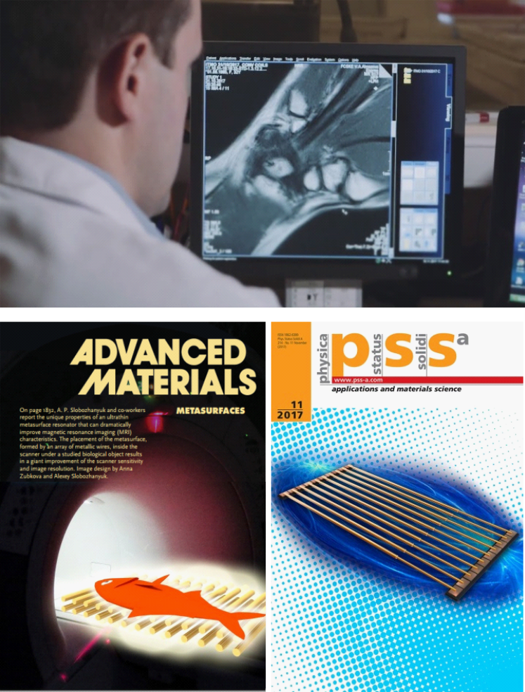
The next logical step was putting this somewhat awkward structure – essentially a bowl of water with brass wires – inside an MRI scanner. As a final touch, the scientists topped the construction with a frozen flatfish purchased at the nearest supermarket. The resulting images were not only bright but also of extremely high quality.

MR scans of the flatfish
“We didn’t understand what was going on, but had already taken our place in the line for a Nobel Prize. Then we finally decided to see for ourselves how MRI scanners actually functioned. Yes, only at that time, having worked with the device for several years. All I can say to our credit is that we believed we had good fundamental education and we knew it all already,” admits Alexey Slobozhanyuk.

Alexey Slobozhanyuk
The reputable journal wasn’t satisfied with an article featuring no experiments on life subjects – and even a follow-up experiment involving rats wasn’t impressive enough for publication. Instead, the article appeared in Advanced Materials, a journal with a slightly lower rating, where it was reviewed by Professor Andrew Webb, who grew interested in the study and offered his help. At his lab in Leiden, Prof. Webb developed surfaces with high dielectric permittivity that produced an effect similar to that acquired by ITMO University researchers. That was the start of a long-standing partnership that continues to this day.

The researchers headed to Europe to fill the gaps in their understanding: first for a seminar in Belgium and then to a lab of their colleagues from Utrecht. There, the whole collective reproduced the experiment with a bowl and a flatfish, while also receiving a professional explanation of what exactly happened inside the scanner to account for such results. These proceedings were described in an article and sent to Nature Materials for review.
The experiment with the reorganizing metasurface
2016-2018: from a bowl to a platform
Now researchers had to make the structure more convenient for medical applications. That’s when Alena Shchelokova (back then, a PhD student and now a research associate at the School of Physics and Engineering) joined the research team.

“Basically, the structure focuses a field around itself and this way, significantly enhances the magnetic resonance signal and the quality of the resulting images. In order to make the wires resonant, we have to set the half-wave resonance at 64 MHz. This would require wires that are several meters long and are thus hard to place inside the machine. The solution is to place the wires in water – thanks to this, their electrical length can be much shorter,” says Alena Shchelokova.

In short, water allows the device to be more compact. Thanks to calculations and optimization, the scientists managed to replace the bowl with a small box filled with water. It’s more suitable for practical use, which was proven by the first volunteer – Prof. Webb himself, who scanned his hand.

Why was there a need for all these bowls, water, and wires in the first place? The thing is, MRI is based on resonance and a bowl with wires is a resonator in itself. As long as its frequency corresponds with that of the scanner, the resonances intersect and enhance the signal.


Alena Shchelokova
MRI scan of Prof. Webb's hand
However, it was clear that the water had to go. Stanislav Glybovsky, a specialist in antennas and radiophysics, joined the team and suggested a new version of the device. It was still based on wires but they could be made shorter thanks to a special parallel-plane condensator. This made the device look like a platform with wires and patches printed on it.


“This was an important milestone, because at that point, there were several radiofrequency devices that could produce high-quality images of high quality on the market. We wanted to compete with them and we did: our device’s characteristics were equally good but it was also safer to use,” says Alena Shchelokova.
Stanislav Glybovsky
2019-2020: from metamaterial plates to ceramic cylinders
Planar structures, however, have one downside: within them an electromagnetic field decays more rapidly, thus making it a challenge to scan anything thicker than a hand.


The developed coils resembled a hollow cylinder assembled from a stack of ceramic rings. As noted by the researchers, this cylinder-like shape instantly determined its further application, namely, breast MRI.
“Creating a wireless MRI device for the mammary gland precisely seemed a promising and crucial task. The reason is, hospitals don’t have enough coils for such procedures, which is especially notable in Russia. On top of that, such coils are rather expensive – not every hospital would purchase this specialized equipment even though breast cancer remains a major public health concern globally,” recalls Alena Shchelokova.
To solve this problem, the scientists turned to a new generation of ceramics-based coils developed by Ceramics JSC, a partner of ITMO’s Faculty of Physics. This type of material possesses higher permittivity, which makes it possible to concentrate electromagnetic energy therein, and super low energy loss sufficient for keeping the field from fading too soon.

At this stage of the project, the scientists started working with volunteers. Clinical trials were performed under the aegis of the Almazov National Medical Research Centre. However, it quickly became clear that ceramics don’t make a great fit as the material is not only rigid but also fragile, heavy, and expensive. Moreover, any scratches or cracks on the surface would significantly affect its properties.
2020-2021: MetaCoil with a Tesla-like body
The researchers had to return to wire resonators, however, this time they decided to place them differently. Given the past experience and the chosen application, they placed the measuring points on two parallelepipeds. The resonators were proven to be reliable: they efficiently focused the field on the required area and produced bright and clear images. Nevertheless, the scientists kept seeking a way to make the technology not only convenient for patients but also visually appealing.

A receive coil that fits inside the MetaCoil device
“We planned to present our technology to our potential clients and partners and were a bit worried about its appearance. We wondered how to make it produce the impression compared to that from a new Tesla. Plus, we still had to figure out how to make it convenient, too. We had already launched clinical trials. Volunteers had to lie in the scanner for 40 minutes and sometimes even hours. It was hard. I remember how we used my measurements to manually create the first body for our device from expanded polystyrene; then we somehow puttied and painted it. The prototype turned out to be inconvenient, rigid, and heavy. But the main problem was that we had no background in design and product development,” says Polina Petrova, manager of the MetaCoil project and innovation commercialization specialist at ITMO’s School of Physics and Engineering.

As a result, the scientists outsourced the task: a Moscow-based company developed the design, made 3D renders, and selected the materials. The body of the device was 3D-printed and its pillows were made of artificial leather and polyester. According to the developers, the current device is much closer to a Tesla in its looks yet it is still a work in progress.

Polina Petrova
What’s next?
The developers of the new MRI device have been working in close cooperation with medical specialists from the start of the project in 2015. Apart from the scientists, research also involves radiologists from the Almazov National Medical Research Centre. Together, they not only test the device but also adapt it to their needs and goals. The developers constantly make the adjustments needed to satisfy the requests of medical specialists. At the moment, the researchers are trying to introduce axillary scanning into their device to detect hidden metastases and lymph node inflammation.
According to Aleksandr Efimtsev, a radiologist at the Centre, the existing prototype is already superior to and more efficient than other models created by major manufacturers. The scientists, however, will have to deal with several issues before introducing their technology into practice.

“Our device can already be used for research but, unfortunately, it doesn’t work for all women yet because of certain anatomical features. We have to find a way to make it suitable for all sizes. Apart from that, there are also some problems with setting up pulse sequences for scanning but it is more about the external design than the coils. They work perfectly and require no changes.
The technology offers numerous advantages. To begin with, it doesn’t weigh much (which is a crucial factor, too) and requires no electrical outlet. As no wires are needed, it’s also mobile and robust. Unlike other coils, the device greatly reduces the number of heart rate artifacts and in general, is more focused on mammary glands than on the entire chest,” explains Aleksandr Efimtsev.
Aleksandr Efimtsev
The scientists continue to refine their product. In particular, they slightly changed the inner structure of the device to a square-based one by combining the original resonator with a new one. They are also working on the optimization of the device’s body and making it suitable for people of different shapes.

Viktor Puchnin, MetaCoil development engineer
Created by:
Text: Ekaterina Shevyreva
Photo and video: ITMO’s School of Physics and Engineering, Lilia Kichigina, Dmitry Grigoriev / ITMO.NEWS
Layout: Ekaterina Shevyreva
Editors: Anna Kirillova, Elena Menshikova
Translators: Kseniia Tereshchenko, Catherine Zavodova, Marina Belyaeva
Text: Ekaterina Shevyreva
Photo and video: ITMO’s School of Physics and Engineering, Lilia Kichigina, Dmitry Grigoriev / ITMO.NEWS
Layout: Ekaterina Shevyreva
Editors: Anna Kirillova, Elena Menshikova
Translators: Kseniia Tereshchenko, Catherine Zavodova, Marina Belyaeva